The addition of CT complements the other specialist imaging technology we have at Oakham; MRI and Scintigraphy and means we now offer the complete range of advanced imaging modalities.
With our specifically designed facility at Oakham we are able to image equine patients’ heads, necks and limbs. The region and size of the horse determines whether or not the imaging can be performed with standing sedation or requires a general anaesthetic.
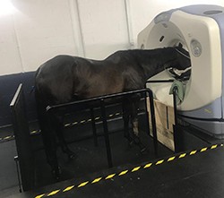 Imaging of the head and upper neck is usually performed with standing sedation. CT is particularly useful for the investigation of problems of the head, since the three dimensional structure of the head makes interpreting x-rays very difficult. We use CT to image dental disease and other causes of nasal discharge or bleeding. It is also possible to evaluate the joints of the jaw (temporomandibular joint), which is relevant in horses that suffer from head shaking. For standing CT the horse stands on a specially designed hydraulic platform which takes the patient through the gantry. The image capture, or scan, takes no longer than a couple of minutes.
The imaging of limbs and the base of the neck requires the horse to have a short general anaesthetic. CT is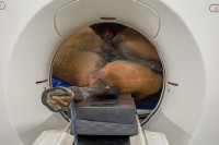 becoming widely used for pre-operative imaging and accurate surgical planning. The 3-D reconstructions allow us to fully understand the pathology, its location and the precise treatment required. In many circumstances the imaging may show that the patient doesn’t require surgery and thus only requires a very short anaesthetic.
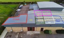 Our CT suite at Oakham is deliberately located adjacent to our operating theatres. Therefore, with pre-operative imaging, CT at Oakham adds very little to total anaesthesia time of patient, whilst ensuring our surgical approach is as accurate and efficient as possible.