Our scintigraphy suite, built in partnership with Nottingham University has been in use since January 2009.
Scintigraphy is also known as ‘Bone Scanning' and is an important diagnostic aid for the examination of lame or poorly performing horses. Common uses would be when:-
- An area of lameness has been localised with the use of nerve blocks but there have been no abnormalities detected on radiography or ultrasonography of the area
- Nerve blocks have not revealed the cause of lameness
- There is multi-limb lameness
- An area cannot be easily penetrated by X-ray (e.g. the pelvis)
The process begins with the injection of a radioactive isotope (technetium 99) into the horse's blood stream,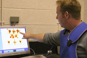 approximately 2 hours before the scan, to allow sufficient time for uptake into the bone. Areas of damage have a greater uptake of the radioactive isotope and when scanned using a gamma camera appear on the image as 'hot spots'. These 'hot spots' highlight areas of bone that are metabolically active, usually indicating a disease process.
Scintigraphy can be very helpful in picking up problems early in the disease process or for identifying suspected fractures. Scintigraphy is typically used in the investigation of subtle lameness of the back, pelvis and upper limbs where the large muscle mass can make nerve blocks, radiography and ultrasound investigation particularly difficult.
As a result of the use of radioactive material, there are very strict protocols that limit the exposure of staff to radiation. Once the horse has been scanned, it is returned to a stable where the radioactivity is able to safely decay, and may not be removed for the next 48 hours. During this time it may not be mucked out, but can receive food, water and hay. Once the horse has been removed from isolation it is common for it to undergo further examination.