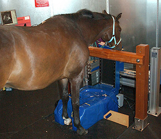
Oakham Veterinary Hospital is one of the few practices in the country with a standing MRI (magnetic resonance imaging) facility.
MRI is a diagnostic imaging technique that involves placing the part of the body to be imaged inside a strong magnetic field. Pulses of radio waves are applied to the area, and a signal (also in the form of radio waves) is received by a computer which generates the image.
The design of the MRI suite is such that scanning can be performed up to the knee and hock region under sedation, without the need for a general anaesthetic. The front shoes (or hind shoes in some cases) must be removed and the whole process can take anywhere between 1 and 3 hours, depending on the size of the area being scanned, and most importantly, the temperament of the patient.
Unlike other imaging techniques, MRI allows the evaluation of both bone and soft tissues at the same time. One of the most common uses for MRI is to assess lameness issues within the hoof capsule, as this can be a difficult area to make an accurate diagnosis. For example, many horses with chronic lameness affecting one or both feet have been found to be suffering from damage to the deep digital flexor tendon (DDFT) or one of the many other ligaments in the foot. MRI is also particularly useful in diagnosing bone diseases such as navicular and pedal bone diseases.